Author: art sci team

  • Project 6: Randomness

    Project 6: Randomness

    Randomness, the lack of a definite pattern or predictability, is usually undesirable in scientific inquiries. Often it poses a nuisance to be overcome by repeated measurements and statistical analysis; in some cases (e.g., at atomic and subatomic scales governed by quantum mechanics), it is an essential property of the system, setting an insurmountable limit to the predictive power of our theory. Nevertheless, randomness in the genetic machinery of neurons has been ingeniously exploited with stunning effects. 

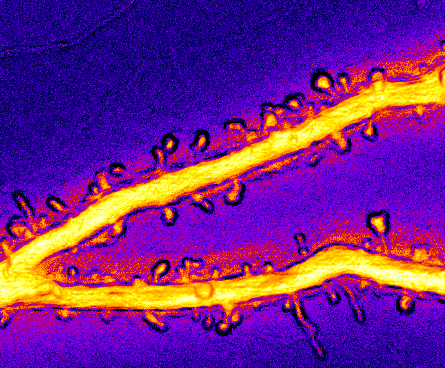
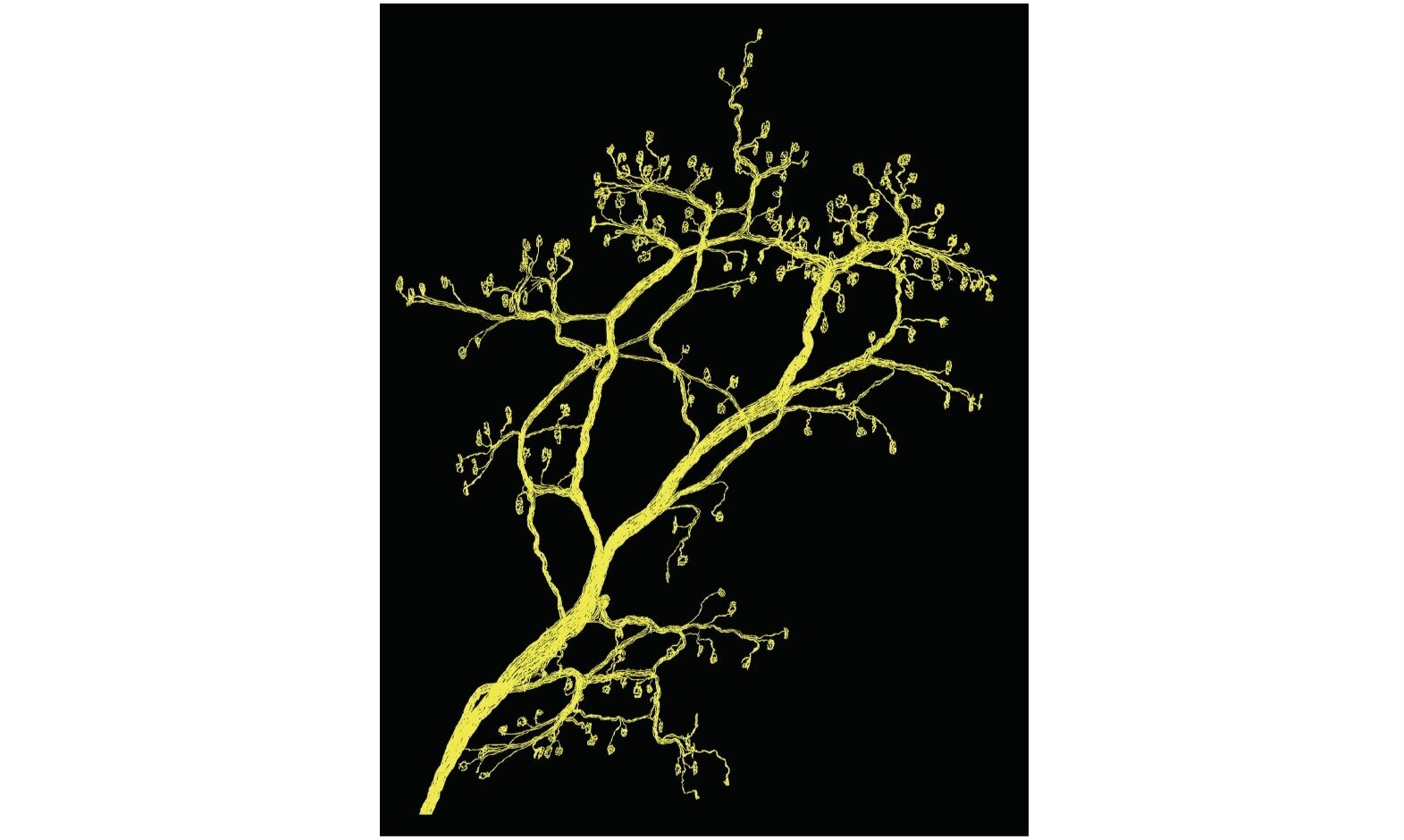
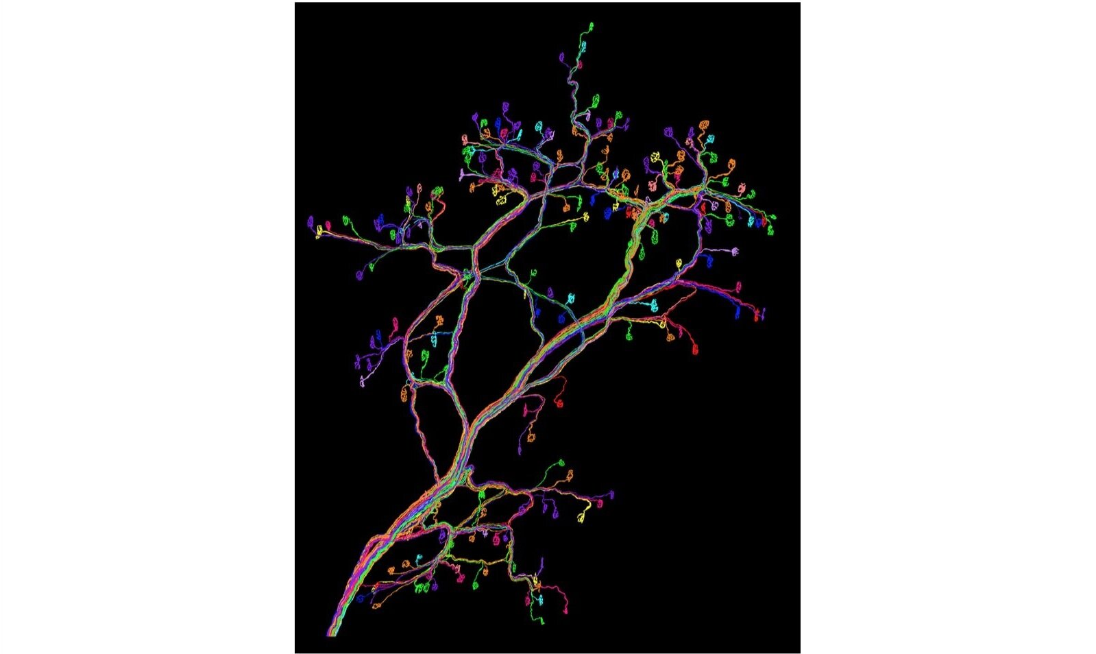
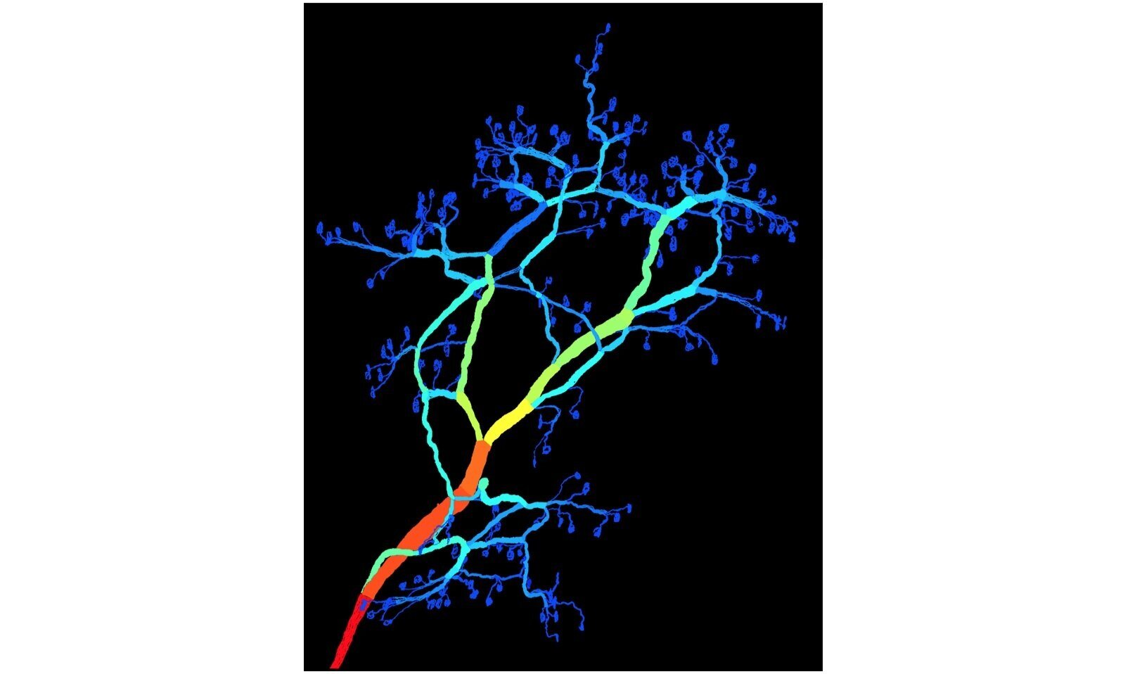
    Brainbow labeling of neurons

    Fluorescent proteins have revolutionized biology, as they enable researchers to visualize cells and tissues of interest with specific labeling. However, each “cell type”, defined by a common pattern of gene expression, comprises many cells, which are often packed too densely to be visualized clearly. Labeling each cell with a distinct color would solve this problem, but there are simply not enough variants in the palette of fluorescent proteins to do so.

    To solve this conundrum, a group of neuroscientists led by Dr. Jeff Lichtman at Harvard University came up with an ingenious idea. They realized that one only needs three kinds of fluorescent proteins to generate many colors, just as the three primary colors (red, green, and blue) can be mixed to yield millions of colors in movies and televisions. However, as cells belonging to the same cell type share the same gene expression pattern, how can they be coaxed into expressing these fluorescent proteins in different proportions?

    The researchers took advantage of the intrinsic randomness of genetic machinery. They made DNA constructs encoding different fluorescent proteins (for example, red, yellow, and cyan) and inserted them into the mouse genome. The expression of these proteins may be turned on or off, depending on the action of an enzyme (Cre recombinase). Notably, the transgenic mouse genome encodes multiple copies of each fluorescent protein, but whether a particular copy is expressed or not is random in any cell. The net effect is that each cell expresses a distinct mixture of red, green, and blue fluorescent proteins and exhibits a unique color.     

    Randomness in art

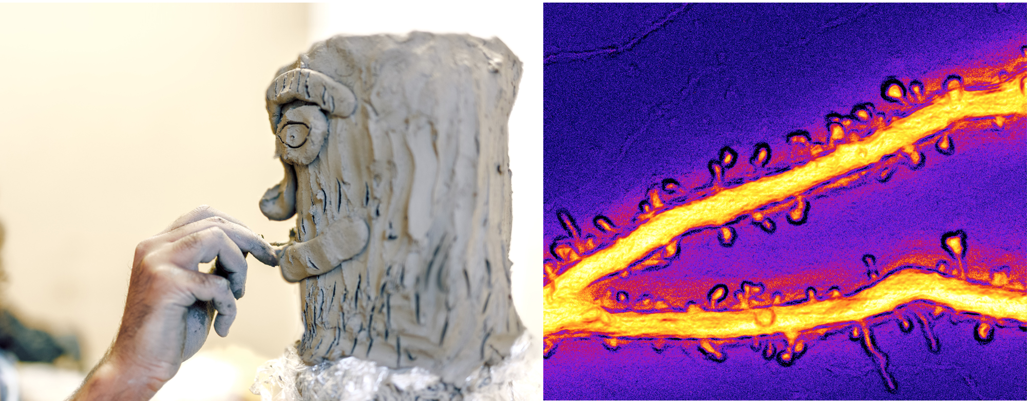
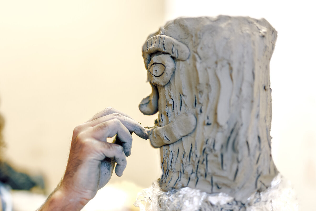
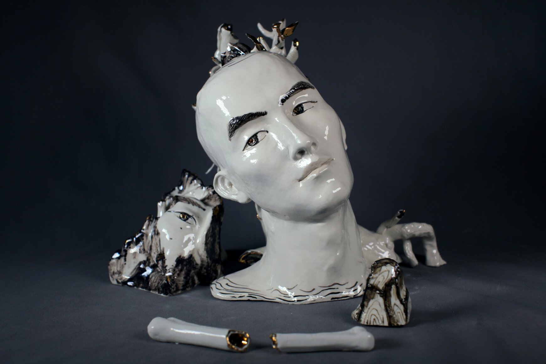
    Artmaking is a deliberate process. We typically envisage an artist judiciously materializing a vision into a physical form – chiseling a piece of marble into a sculpture or meticulously rendering a three dimensional horizon onto a two dimensional canvas. This view of art creation emphasizes the artist’s intentional, conscious control over the materials to create an aesthetic object. Randomness, on the other hand, is by definition unpredictable and uncontrollable. It seems to be at odds with the purposefulness of artmaking. Paradoxically, many artists deliberately embrace randomness either as a medium itself or as an essential ingredient of the final product.

    Jackson Pollock, the abstract expressionist painter, for instance, is famous for his “drip” paintings. By splattering paint onto the canvas, Pollock introduced an element of chance into his work. While his actions were deliberate, the outcome was inherently unpredictable. Once the paint left the brush, where it would land on the canvas was no longer controlled by the artist’s hand. The random outcome resulted in surprising, often unplanned effects.

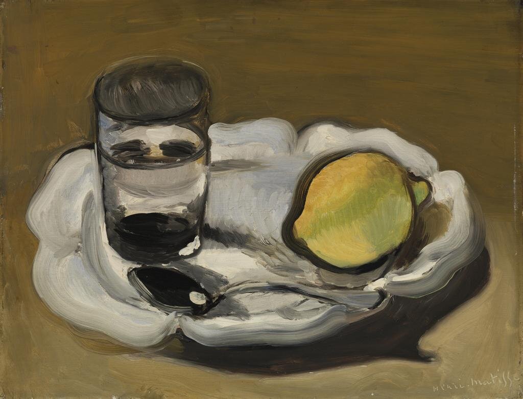
    In other cases, randomness is not merely a tool to facilitate art creation but a central component of the artwork itself. For instance, Fred Whipple, an American astronomer, created a series of “stochastic paintings,” which he defined as compositions created through random processes. He asked whether creativity, self-expression, or beauty could emerge from randomness. He proposed that stochastic art should follow rules governing randomness. Importantly, Whipple distinguished between randomness and mere irregularity. He explained that within an aggregate of random numbers, colors, or distributions, structured patterns would naturally arise and lead to something that appears ordered, not mere chaos. To create his paintings, Whipple selected random numbers and applied a set of rules to choose shapes, shades, and colors, embracing randomness to yield structured yet unpredictable compositions.

    Jean Arp, a Dadaist painter, sculptor, and poet, famously incorporated chance into his art by tearing up pieces of his own drawings and allowing them to fall onto the canvas, preserving their landing spots. He was among the first artists to use randomness as a fundamental element of his work, treating chance as a collaborator rather than a limitation. By relinquishing control, Arp challenged the notion that artistic skill required precision and intent, instead embracing unpredictability as part of his creative process.

  • Project 5: Carpe Tempus

    Project 5: Carpe Tempus

    Two kinds of events evade the naked eye: those happening too slowly to be noticeable, and those happening too fast to be discerned. While a natural process usually cannot be accelerated for observation, technology has enabled people to catch the moment when a fast process occurs and record it as impressive images.


    Science Work

    One neuron transmits information to another cell by releasing chemicals at specialized sites called synapses. The synapse consists of two parts: the presynaptic part on the sender neuron and the postsynaptic part on the recipient cell. The chemicals (“neurotransmitters”) are stored in small lipid packets called vesicles, residing in the presynaptic site. When the sender neuron generates an electrical pulse, it triggers the rapid release of neurotransmitters, which bind to receptor proteins on the postsynaptic site and causes the recipient cell to respond.  The mechanism of this process is a central question in neurophysiology. However, as releasing is very fast (on the order of milliseconds), it is difficult to capture it in action. 

    In the early 1970s, two American scientists, Tom Reese and John Heuser, designed an ingenious device to capture the moment of neurotransmitter release (Figure 1). They placed a piece of frog muscle with the nerve on a “freezing head”, which can free-fall onto a block of copper cooled by liquid helium (-269 °C or -452.20 °F). During the fall, the nerve is electrically stimulated by wires in the freezing head. By the time the neurotransmitters are released, the tissue is in contact with the cooling block and gets frozen instantaneously. The vesicles at different stages of fusing with the presynaptic membrane and opening up to release their neurotransmitter contents can thus be captured on electron microscopic images (Figure 2, top), from which one can reconstruct the likely sequence of events (Figure 2, bottom). This works provides a definitive support to the prevailing model of how neurotransmitter release occurs.

    References: 
    Heuser, J.E., et al. (1979) “Synaptic vesicle exocytosis captured by quick freezing and correlated with quantal transmitter release.” J. Cell Biol. 81: 275-300 

    Heuser, J.E. and Reese, T.S. (1981) “Structural changes after transmitter release at the frog neuromuscular junction.” J. Cell Biol.88: 564-80

    Artwork

    Art can turn an ephemeral event into an object with eternal beauty. This is amply demonstrated by the examples below, in which visual artists, employing clever technologies, freeze events that are too fast for the naked eye to appreciate. 

    Figure 1: Bullet through Apple

    This striking image captures the moment when an apple is pierced by a bullet. The duration of the flash here is ⅓ of a microsecond; the room is otherwise dark. According to the MIT Museum, “to trigger the flash at the proper moment, a microphone, placed a little before the apple, picks up the sound from the rifle shot, relays it through an electronic delay circuit, and then fires the microflash.”

    Figures 2.1 and 2.2: Water Sculptures

    When a water droplet collides with the surface of a water pool, it creates a splash with exquisite form that only lasts a tiny fraction of a second before collapsing. To freeze the liquid in motion, artists mix water with other materials such as guar gum and dyes to make it denser and colorful, and use high speed photography to capture the fascinating moment when the “liquid sculptures” emerge. 

  • Project 4: Yin and Yang

    Project 4: Yin and Yang

    Scientific images usually have a clear distinction between the object of interest and the background. Classical painting adopts a similar perspective between the foreground and the background. However, such distinctions are in a sense artificial, as they depend on where the viewer’s attention is accorded. The illustrations given below abandon this dichotomy; instead they leverage and emphasize the symmetry and complementarity between the “foreground” and the“background”, as the Yin and Yang in Taoist philosophy.


    Science Work

    The brain is packed with nerve cells. Each nerve cell has many extricate processes that may be very thin and highly tortuous. Together, this dense network of subcellular structures surpasses the resolving power of conventional optical microscopes. Moreover, all cells in the brain are surrounded by extracellular space (ECS), an exquisite labyrinth filled with fluid. The caliber of “tunnels” and “canals” of ECS also falls below the resolution limit of conventional optical microscopes. To observe the nanoscale organization of the neuronal network or, conversely, that of the ECS, Dr. U. Valentin Nägerl and colleagues devised SUSHI (super-resolution shadow imaging), which cleverly uses negative images. They perfuse the ECS with a fluorescently labeled fluid and imaged it with STED microscopy (a Nobel prize-winning super-resolution technique developed by Dr. Stefan Hell). The cell bodies and processes, unlabeled by the fluorescent dye, become dark voids in the image. Therefore, inverting the Lookup Table (LUT) of the raw image yields a clear image of the cellular structures (Figure 1). Moreover, if a cell is labeled with a fluorescent molecule of a distinct color, the superimposed positive image of the cell (yellow) and the inverted image of ECS (gray) together give the spatial organization of processes of cells that contact it, either sending or receiving information from it (Figure 2).

    Ref: Jan Tønnesen et al., Super-resolution imaging of the extracellular space in living brain tissue. Cell 172: 1108-1121, 2018.  

    Artwork

    Traditionally, visual artists portray important objects in the foreground, relegating less important ones to the background.  The contrast between the two naturally directs the viewer’s attention to the intended focal subject.   

    However, in Untitled (Stairs) (Figure 1), the artist Rachel Whiteread swaps the concreteness of a familiar object with the emptiness that we normally associate with non-being, by creating a monumental plaster cast of the space surrounding a staircase.  As stated by The Tate Modern’s description:

    “Whiteread’s casting process has transformed the stairs into an abstracted geometric composition which combines physical familiarity with a mental conundrum – that of trying to envisage the original structure from which the new object has been derived.”

    Traditional Chinese landscape painting makes clever use of blank space (Figure 2).  Intentionally left on the paper, the blankness may represent the sky, the river, rolling fields, etc. and thus acquires a concrete meaning in the context.  The calculated arrangement of “foreground” objects in the painting cues the viewer to substantiate the blank space with his or her own imagination. 

    Nullifying the usual distinction between foreground and background leads to images that evoke unconventional viewing experiences. The famous Dutch graphic artist Maurits Cornelis Escher and the Cuban-American abstract painter Carmen Herrera tease our perception by interdigitating two sets of images (Figures 3 and 4). Such juxtaposition forces the viewer’s brain to flip continually between two mutually incompatible interpretations: Is the image black fish and birds with a white background, or white fish and birds with a black background? Is it an orange comb on a green background, or a green comb on an orange background? 

  • Project 3: The Sediments of Time

    Project 3: The Sediments of Time

    Life unfolds as a continuum of events in time. A single snapshot, be it a photo, a painting, or a sculpture, may capture a particular event vividly, but it is not straightforward to render a dynamic process in a single image.  Both neuroscientists and artists have devised special techniques to crystalize a prolonged process into a single picture. 


    Science Work

    Neurons process and transmit information by firing electrical spikes called action potentials. To decipher how the brain works, we would ideally record all such electrical activities of each neuron continuously. This lofty goal is still beyond the reach of current technology. Taking a step back, neuroscientists have come up with clever ways – either by leveraging biological processes or via protein engineering – to condense the history of neuronal activities into single-snapshot readouts. 

    When neurons are activated, the so-called immediate-early genes are turned on transiently. Therefore, if a neuron has a high level of the protein products of such genes, it is likely to have been active recently. Figure 1 shows an example of mouse brain neurons activated by a single session of stress as revealed by staining the protein product of an immediate-early gene called c-Fos. Clearly, stress activates a lot of neurons! 

    Neuronal activity is also accompanied by an influx of calcium ions into the cell. Recently, researchers have engineered a fluorescent protein called CaMPARI, which changes color from green to red as it binds to calcium. The more active the neuron is, the more calcium ions flow in, and the redder the neuron becomes. Figure 2 shows how the zebrafish brain labeled with CaMPARI changes color under different conditions. For example, the anesthetized brain is quiet; swimming activates certain brain regions; seizure, heat, or cold exposure activates more cells but again with distinct patterns (images courtesy of Dr. Eric Schreiter; see Fosque, B. F. et al., Labeling of active neural circuits in vivo with designed calcium integrators. Science 347: 755–760, 2015). 

    Artwork

    Artmaking is rooted in one of the most innate human desires – to capture and depict histories by images.  From cave wall murals, to Chinese calligraphy and ink wash paintings, to the Impressionists’ depictions of light, artists attempt to represent undefinable movement over time – an event, an essence, an emotion – as a single visual image, ultimately with the hope of telling a story that outlasts human life.

    As technology progresses, tools to capture these processes become more efficient, immediate, and accessible.  Artists thus begin to push the limits of these tools to fulfill the desire of capturing history with a single image. For example, many photographers creatively use long-exposure photography, opening the camera shutter over many seconds, minutes, hours, or even days, to overlay a series of moments in one exposed image.  While many details may be blurred out, this technique gives the viewer a holistic view of the process, perhaps bringing out the intrinsic dynamism better than a photograph that presents just a snapshot at a single moment.

  • Project 2: Plus and Minus

    Project 2: Plus and Minus

    A sophisticated masterpiece is never made overnight. It takes shape in a long process of adding new materials, removing undesired ones, or both. The same principle applies to artworks and to the maturation of the nervous system.


    Scientific Illustrations: changing neural circuit

    The human brain consists of billions of nerve cells (neurons). Neurons connect with each other at specialized sites called “synapses”, a term coined from two Greek words meaning “to fasten” and “together”. As each neuron may connect with thousands of other neurons, the resulting network is incredibly complex. Moreover, this network is not static. New synapses are constantly formed, and some old synapses are gradually removed. This process is further influenced by experiences such as sensory inputs, motor exercises, and the reward or punishment derived from actions, so it continuously shapes the structure of the brain circuit to make us better adapt to the environment.

    A glimpse into the changing neural circuit. We repeatedly imaged the same dendrite (the input cable of a neuron) and the dendritic spines (the little protrusions from the dendrite, which are the sites where synapses reside) in the brain of a living mouse. While the dendrite remains unchanged, dendritic spines emerge and disappear over time, representing the remodeling of connections between neurons in the neural circuit.

    Artworks: Clay Modeling

    Many familiar artistic practices involve adding materials. Oil painting, watercolor, and traditional Chinese painting apply ink or pigments atop a substrate (canvas or paper). Other practices require removing materials to create the desired outcome: imagine chipping away at a marble to make a statue. 

    In clay modeling, however, both addition and subtraction are integrated in a fluid process that culminates in the final product, just as the creation and elimination of neural connections are both indispensable for the brain to learn and to adapt to the  environment.

    The artist starts with a piece (or pieces) of clay and shapes it into a rough form. Then he or she begins to carve, peel, remove, or smooth (all “subtractive” processes) it into a detailed object; at the same time, more clay may be added. This process is repeated until the clay finally transforms into a refined artwork  as the artist envisions it.

  • Project 1: Depicting the Real

    Project 1: Depicting the Real

    Reality has many facets.  It is almost a self-evident truth in the world of arts, as each artist can choose to depict the same object from his or her unique viewpoint.  Less appreciated is the fact that a scientific image also represents an aspect of the object that interests the investigator, rather than what the object “really looks like.”  


    Scientific Illustrations: the neuromuscular circuit

    Here are three images of the same object: the processes of all nerve cells (“neurons”) that project into, and form contacts with, a small muscle. These neurons are filled with a yellow fluorescent protein, so they are visible under a fluorescent microscope.  Each of these neurons sends out a long process called the axon, which is the output cable of the neuron to convey electrical signals to its targets. Once it reaches the target muscle, the axon branches out like a tree. The flower-like structure at the end of each twig is the contact with a single muscle fiber, called the neuromuscular junction, through which the neuron commands the contraction of the muscle fiber.  In the adult muscle, each neuron controls numerous muscle fibers, but each muscle fiber receives input from only one neuron.  

    Image 1 is close to what this neuromuscular circuit appears to the naked eye under the fluorescent microscope – a collection of glowing spaghetti. But even this image omits many elements, such as the muscle fibers and other types of cells surrounding the junctions: we chose to label only the neurons.

    Image 2 is the outcome of laborious image analysis.  Each neuron is rendered in a distinct color, so its branches are visually disentangled from those of other neurons.  Now we can easily tell which neuron controls which muscle fiber – the circuit diagram is revealed. 

    Image 3 highlights the collective behavior of axons.  Imagine that the nerve bundle is a river, which branches out into smaller and smaller tributaries (branches of axons) until each terminates in a reservoir (neuromuscular junction).  Imagine also that the water volume in each tributary is proportional to the number of terminal reservoirs it supplies.  The colors represent the water volume in each segment of the tributaries.  In other words, a reddish color means more neuromuscular junctions downstream of the segment. 

    Which of the three images is “true”? None, as each emphasizes one aspect of the neural circuit at the cost of others; all, as each is a faithful representation of the things we chose to focus on.    

    Ref: Adapted from Lu J et al. (2009) The Interscutularis Muscle Connectome. PLoS Biology 7(2): e1000032. https://doi.org/10.1371/journal.pbio.1000032

    Artworks: Still life

    The three still life paintings are executed by different artists throughout three distinct periods of art history. Although they depict similar objects (lemon, pitcher, dish-ware), the choice of artistic focus and techniques differ dramatically. As seen in these three chronological images, representation becomes more and more abstracted as the timeline of canonical art history progresses. Does this mean any one image is more or less ‘real’ than the other? Some might argue yes, some might argue quite the opposite. Through highlighting techniques like color, light, form, and spatial relationship, the artists are presenting the same objects to the viewer in different ways, evoking specific but varied aesthetic and emotional responses.

    Artwork 1: A member of the Dutch Golden Age, Willem Kalf was one of the great still-life painters of the seventeenth century. True to Kalf’s style, this image intentionally portrays rare objects that appealed to the wealthy Dutch class, organized very precisely against a dark background. Kalf uses light, sheen, and rich textures to convey the luxury of the objects to the viewer. Despite the hyper-realistic painting style, the intentionally staged scene is in a sense, a false representation of reality.

    Artwork 2: Henri Matisse, known as the greatest colorist of the 20th century, used color, outline, and flattened forms to depict a scene on a canvas. Matisse used intentional relationship between form, space, and color to make objects appear both at once floating in space and situated realistically in the pictorial plane. Here, Matisse uses muted colors to show highlights and lowlights, and to ultimately bring the scene to life.

    Artwork 3: Roy Lichtenstein, considered one of the most seminal American pop artists, references the techniques of the Cubists in this hybrid piece. Cubists (most notably Pablo Picasso and Georges Braque) were interested in depicting the “real,” but not in the traditional style of previous eras. Historically, artists used optical illusions like linear perspective, single vanishing points, and shading to turn the two-dimensional plane into a three-dimensional scene. Cubists rejected the idea that art had to precisely mimic nature. Cubists, instead, used fractured and geometric shapes and multiple vanishing points to emphasize the two-dimensional nature of the canvas, but suggest the three-dimensional quality of objects.