Project 5: Carpe Tempus
Two kinds of events evade the naked eye: those happening too slowly to be noticeable, and those happening too fast to be discerned. While a natural process usually cannot be accelerated for observation, technology has enabled people to catch the moment when a fast process occurs and record it as impressive images.
Science Work
One neuron transmits information to another cell by releasing chemicals at specialized sites called synapses. The synapse consists of two parts: the presynaptic part on the sender neuron and the postsynaptic part on the recipient cell. The chemicals (“neurotransmitters”) are stored in small lipid packets called vesicles, residing in the presynaptic site. When the sender neuron generates an electrical pulse, it triggers the rapid release of neurotransmitters, which bind to receptor proteins on the postsynaptic site and causes the recipient cell to respond. The mechanism of this process is a central question in neurophysiology. However, as releasing is very fast (on the order of milliseconds), it is difficult to capture it in action.
In the early 1970s, two American scientists, Tom Reese and John Heuser, designed an ingenious device to capture the moment of neurotransmitter release (Figure 1). They placed a piece of frog muscle with the nerve on a “freezing head”, which can free-fall onto a block of copper cooled by liquid helium (-269 °C or -452.20 °F). During the fall, the nerve is electrically stimulated by wires in the freezing head. By the time the neurotransmitters are released, the tissue is in contact with the cooling block and gets frozen instantaneously. The vesicles at different stages of fusing with the presynaptic membrane and opening up to release their neurotransmitter contents can thus be captured on electron microscopic images (Figure 2, top), from which one can reconstruct the likely sequence of events (Figure 2, bottom). This works provides a definitive support to the prevailing model of how neurotransmitter release occurs.
References:
Heuser, J.E., et al. (1979) “Synaptic vesicle exocytosis captured by quick freezing and correlated with quantal transmitter release.” J. Cell Biol. 81: 275-300
Heuser, J.E. and Reese, T.S. (1981) “Structural changes after transmitter release at the frog neuromuscular junction.” J. Cell Biol.88: 564-80
Artwork
Art can turn an ephemeral event into an object with eternal beauty. This is amply demonstrated by the examples below, in which visual artists, employing clever technologies, freeze events that are too fast for the naked eye to appreciate.
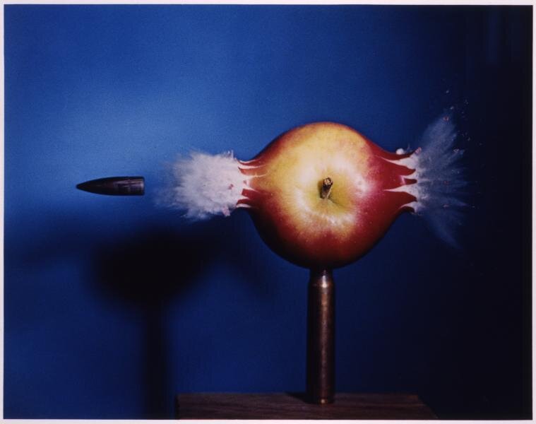
Figure 1: Bullet through Apple
This striking image captures the moment when an apple is pierced by a bullet. The duration of the flash here is ⅓ of a microsecond; the room is otherwise dark. According to the MIT Museum, “to trigger the flash at the proper moment, a microphone, placed a little before the apple, picks up the sound from the rifle shot, relays it through an electronic delay circuit, and then fires the microflash.”
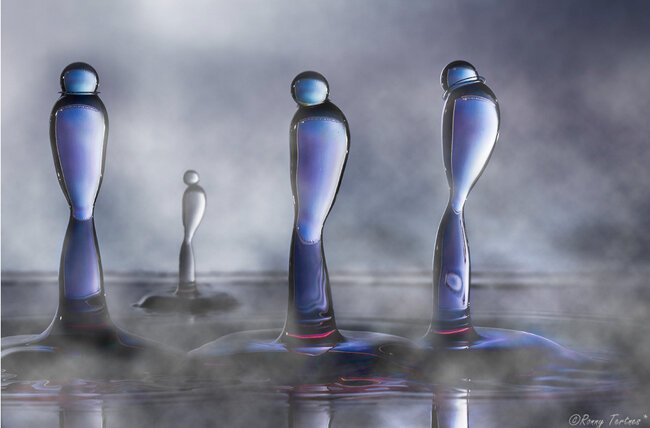
Figures 2.1 and 2.2: Water Sculptures
When a water droplet collides with the surface of a water pool, it creates a splash with exquisite form that only lasts a tiny fraction of a second before collapsing. To freeze the liquid in motion, artists mix water with other materials such as guar gum and dyes to make it denser and colorful, and use high speed photography to capture the fascinating moment when the “liquid sculptures” emerge.


